Nanoparticle Tracking Analysis (NTA) has become one of the most widely adopted techniques for measuring nanoparticle size distribution and concentration in liquid suspension. Its ability to analyze particles individually rather than as an ensemble population provides significant advantages when working with heterogeneous samples such as extracellular vesicles, lipid nanoparticles, viruses, protein aggregates, and polymeric nanomaterials.
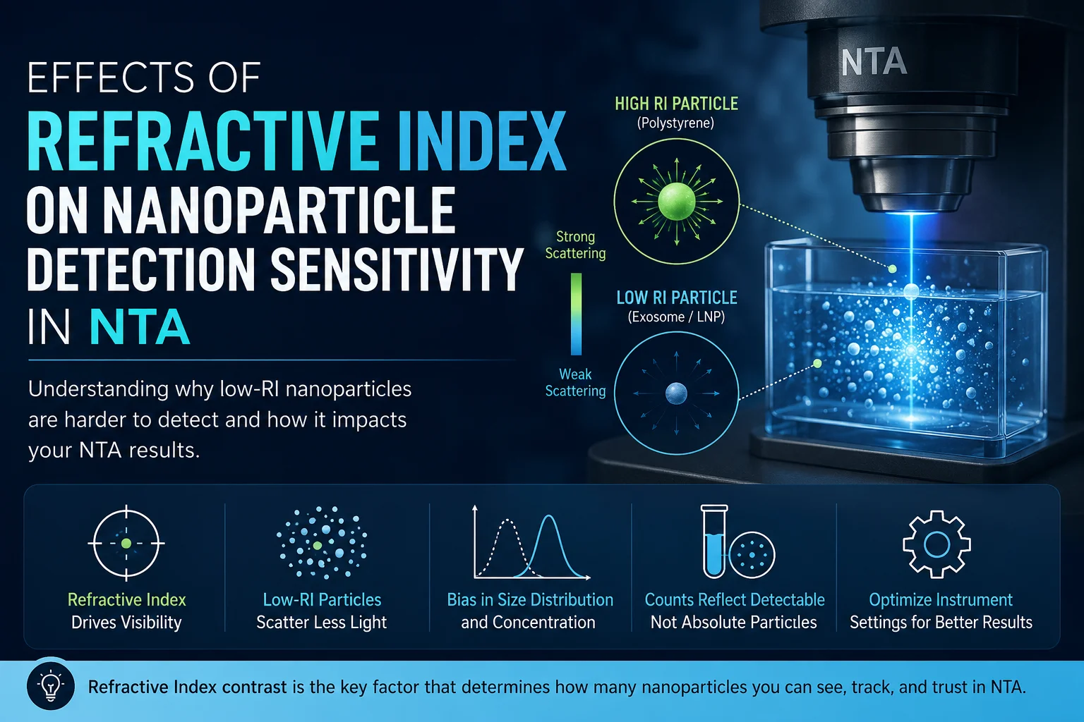
However, one of the most important and often misunderstood factors affecting NTA performance is the refractive index (RI) of the particles being analyzed. Refractive index directly influences how efficiently particles scatter light, which ultimately determines whether particles can be detected, tracked, and accurately measured by the instrument.
For many biological nanoparticles, particularly extracellular vesicles (EVs) and lipid nanoparticles (LNPs), low refractive index presents a major analytical challenge. Researchers frequently encounter reduced sensitivity, undercounting of small particles, or apparent inconsistencies in concentration measurements without fully understanding that refractive index effects may be the underlying cause.
Understanding Refractive Index in Nanoparticle Analysis
Refractive index describes how strongly a material bends or slows light relative to the surrounding medium. In nanoparticle characterization, the refractive index difference between the particle and the surrounding solvent determines the intensity of scattered light detected by the imaging system.
Particles with a large refractive index contrast relative to the surrounding medium scatter light more efficiently and therefore appear brighter during analysis. Conversely, particles with refractive indices close to that of the solvent scatter weakly and become more difficult to detect.
For example:
| Material | Approximate Refractive Index |
| Polystyrene beads | ~1.59 |
| Silica nanoparticles | ~1.45 |
| Water | ~1.33 |
| Lipid nanoparticles | ~1.37–1.42 |
| Extracellular vesicles | ~1.37–1.40 |
| Protein aggregates | ~1.35–1.45 |
Biological nanoparticles typically exhibit much lower refractive indices than calibration beads commonly used during instrument validation. As a result, the practical detection limit for biological samples is often substantially larger than the nominal detection limit advertised for highly scattering polystyrene particles.
Light Scattering and Detection Sensitivity in NTA
NTA detects nanoparticles by illuminating particles with a laser and imaging the scattered light using a sensitive camera. The brightness of each detected particle depends heavily on the amount of light scattered toward the detector.
For small particles operating within the Rayleigh scattering regime, scattering intensity scales approximately with the sixth power of particle diameter:
I∝d^6
This relationship has profound implications for nanoparticle detection. Even small decreases in particle diameter can produce dramatic reductions in scattered intensity.
Refractive index further modifies scattering efficiency. A low-RI particle may scatter orders of magnitude less light than a similarly sized high-RI particle. Consequently, detection sensitivity depends not only on particle size but also on optical contrast between the particle and the surrounding medium.
This explains why:
- 50 nm polystyrene particles may be readily detectable
- 50 nm extracellular vesicles may fall below detection threshold
- concentration measurements may underestimate low-scattering populations
- multimodal distributions may appear artificially shifted toward larger particles
Refractive Index Contrast and Optical Visibility
The critical parameter influencing scattering intensity is refractive index contrast: Δn=n_p-n_m
Where:
- n_p = particle refractive index
- n_m = medium refractive index
As the refractive index difference decreases, scattering intensity declines substantially.
Biological nanoparticles often exist in aqueous buffers with refractive indices close to the particles themselves. This creates inherently weak optical contrast. In contrast, polystyrene calibration beads possess significantly higher refractive indices, generating strong scatter signals even at relatively small diameters.
This discrepancy can create misleading assumptions regarding instrument sensitivity if researchers do not account for refractive index effects during experimental design and data interpretation.
Detection Threshold Bias in Low-RI Samples
Every NTA system uses a detection threshold to distinguish true particle signals from background noise. Particles producing signals below this threshold are effectively invisible to the tracking algorithm.
Low-refractive-index nanoparticles are especially vulnerable to threshold-related bias because their scattered intensity may approach the noise floor of the imaging system.
This produces several common artifacts:
- Undercounting Small Particles: Smaller particles with low scatter intensity may not be detected consistently, causing concentration underestimation.
- Artificial Distribution Shifts: Since larger particles scatter more strongly, the measured size distribution may become biased toward larger diameters.
- Reduced Repeatability: Marginally detectable particles may appear intermittently across measurements, increasing run-to-run variability.
- Loss of Multimodal Resolution: Weakly scattering subpopulations may disappear entirely from measured distributions.
These effects are especially important in extracellular vesicle research, where many vesicles exist near or below the practical optical detection limit.
Biological Nanoparticles Present Unique Challenges
Biological nanoparticles are particularly difficult to analyze because they combine several unfavorable optical properties simultaneously:
- Low refractive index
- Small particle size
- Broad heterogeneity
- Irregular morphology
- Dynamic aggregation behavior
Extracellular vesicles and lipid nanoparticles frequently produce weak scattering signals compared to synthetic nanoparticle standards.
As a result, researchers may incorrectly interpret apparent differences between samples as biological variation when the true cause is altered detection sensitivity arising from optical limitations.
For example:
- EV isolation methods may appear to change concentration
- storage conditions may appear to alter particle distributions
- formulation buffers may influence apparent sizing results
- dilution protocols may affect measured particle counts
In many cases, refractive index effects contribute significantly to these observations.
Refractive Index Effects on Concentration Measurements
NTA concentration measurements rely on counting detectable particles within a defined imaging volume. If low-scattering particles are not detected reliably, measured concentration becomes biased downward.
This problem becomes increasingly severe for:
- small EVs
- low-density liposomes
- dilute biological samples
- weakly fluorescent subpopulations
Importantly, detection efficiency is not uniform across all particle sizes. Smaller low-RI particles are disproportionately underrepresented.
Consequently, measured concentrations should often be interpreted as “optically detectable particle concentration” rather than absolute particle concentration.
Understanding this distinction is essential when comparing:
- different particle types
- different instruments
- different buffer systems
- different laser wavelengths
- different camera settings
Influence of Wavelength and Optical Configuration
Scattering efficiency also depends on laser wavelength. Shorter wavelengths generally improve sensitivity for smaller particles because scattering intensity increases as wavelength decreases.
However, shorter wavelengths may also increase:
- background scattering
- photodamage risk
- fluorescence overlap complications
- solvent noise
Instrument optical design therefore plays a major role in determining practical detection sensitivity for low-refractive-index nanoparticles.
Critical factors include:
- laser power stability
- camera sensitivity
- numerical aperture
- optical alignment
- detection algorithms
- background suppression methods
Advanced systems with optimized imaging sensitivity can significantly improve detection of biologically relevant nanoparticle populations.
Conclusion
Refractive index is one of the most critical factors governing nanoparticle detection sensitivity in Nanoparticle Tracking Analysis. Because scattering intensity depends strongly on optical contrast, low-refractive-index biological nanoparticles frequently present substantial analytical challenges.
Extracellular vesicles, lipid nanoparticles, and other biologically derived particles scatter significantly less light than synthetic calibration standards, often resulting in reduced sensitivity, undercounting, and measurement bias.

Dynamic and innovative sales and product marketing manager with proven success in sales and market share growth in the analytical instruments industry. Combining in-depth analysis, marketing strategy, business process optimization, and “outside the box” solutions to launch new products and revitalize ongoing business.
Introduced NanoSight’s Nanoparticle Tracking Analysis (NTA) technique to the market, a new and significantly advanced method for sizing and counting nanomaterials. Building market recognition and acceptance through marketing initiatives, customer contact, and publication of technical articles.